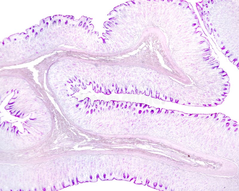
Gastric mucosa. PAS method
Very low magnification light micrograph of the gastric wall stained with PAS method. The inner layer is the mucosa that shows many folds. In the axis of these folds is the submucosa. In the mucosa, the mucous surface epithelium and foveolar cells of gastric pits show a great PAS positivity
View Full Image on Dreamstime
Keywords:
gastricpitmucosabiologydigestivesystemgastrointestinaltracthistologymucouscelllightmicroscopemicrographmicroscopypasmethodstainstomachsubmucosatissue
Username: Jlcalvo
Editorial: No
Width: 3840 pixels
Height: 3072 pixels
Downloads: 0
Image ID: 310913694